Ventricular Tachycardia
The heart is comprised of 4 chambers; 2 atria, which sit on 2 ventricles. An electrical “timekeeper”, called the sino-atrial node, is located at the top of the right atrium. This is the heart’s natural pacemaker, sending out regular electrical impulses that travel through pathways which run down the atria, through the ‘junction box’ of the heart (known as the AV node) and into the ventricles, allowing the heart to contract and relax regularly and pump blood around the body.
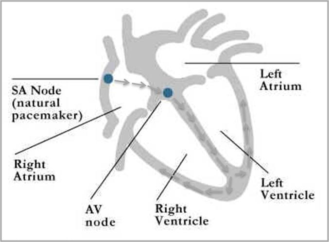
Normal ‘sinus’ rhythm (see text for details)
Ventricular tachycardia (VT) occurs when electrical stimuli are initiated from within the ventricles and then spread through the ventricles, over-riding the normal activity of the sino-atrial node at a much faster rate. These abnormal stimuli cause the heart to beat at over 120-200 beats per minute, in contrast to the normal rate of 60-100 beats per minute. There are different types of VT – some are seen in patients with a completely normal heart and are not life threatening, but some are seen in patients with other forms of heart disease and these can be very serious and even fatal. Symptoms can include palpitations, dizziness, breathlessness; in some instances, also chest pain and blackouts.
Investigation of VT includes initially taking a simple ECG. Sometimes ambulatory ECG monitoring is needed followed by more technical EP studies to identify the site of the electrical impulses. Treatment can be with medication. However, some people benefit from insertion of an Implantable Cardioverter Defibrillator which administers an electrical shock if a life-threatening heart-rate is detected due to the VT. Some patients may also benefit from catheter ablation.